Difference between revisions of "Overview of System Components"
| Line 11: | Line 11: | ||
The depth of imaging is limited to around 200um due to the nature of wide-field fluorescence imaging. As the imaging depth increases, the excitation light intensity decreases due to absorption of the above tissue while the scattering of emission light increase. This becomes more of an issue when imaging regions where labeled cells are overlapping along the z-axis. | The depth of imaging is limited to around 200um due to the nature of wide-field fluorescence imaging. As the imaging depth increases, the excitation light intensity decreases due to absorption of the above tissue while the scattering of emission light increase. This becomes more of an issue when imaging regions where labeled cells are overlapping along the z-axis. | ||
| + | |||
| + | '''Using this system we have successfully imaged Hippocampal CA1, Subiculum, and Visual Cortex using 2mm diameter GRIN lenses from Grintech.''' While thinner GRIN lenses should theoretically be compatible with our system we have limited our initial development to larger lenses due to supply and experimental constraints. | ||
*Specifications | *Specifications | ||
Latest revision as of 14:50, 12 January 2016
The functional design of our microscope is similar to a table top wide-field fluorescence microscope with the following key differences:
- All nonessential components are removed
- The table top objective is replaced with an imaging objective GRadient Index of Refraction (GRIN) lens
- The excitation light source is a super bright LED rather than a lamp or laser
- The optical filters and dichroic mirror are custom cut to minimize their size and weight
- A small CMOS imaging sensor is used to record emission light instead of a large CCD, emCCD, or sCMOS camera
- Adjustment of focal plane is done by moving imaging sensor not objective
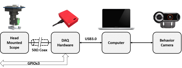
Our miniature fluorescence microscope system consists of a lightweight microscope body, optical components, imaging sensor, and read out electronics. The readout electronics handle all communication between the onboard microscope electronics and the host computer such as LED intensity, imaging sensor configuration, and imaging sensor data. The body is machined out of 3 pieces of black Delrin plastic and assembled using self-threading, 1mm diameter screws. The optical components collimate the excitation light, band-pass filter the excitation and emission light, and focus the emission image onto the imaging sensor.
The depth of imaging is limited to around 200um due to the nature of wide-field fluorescence imaging. As the imaging depth increases, the excitation light intensity decreases due to absorption of the above tissue while the scattering of emission light increase. This becomes more of an issue when imaging regions where labeled cells are overlapping along the z-axis.
Using this system we have successfully imaged Hippocampal CA1, Subiculum, and Visual Cortex using 2mm diameter GRIN lenses from Grintech. While thinner GRIN lenses should theoretically be compatible with our system we have limited our initial development to larger lenses due to supply and experimental constraints.
- Specifications
- 700μm x 450 μm field of view
- 6x to 7x magnification
- ~1um per pixel
- 50μm to 200μm working distance
- <3 grams
- Full-field frame rate up to 60Hz
- Resolution: 752px x 480px
- Super Speed USB: 6Gb/s
- Commutator compatable
Details on specific components of the system can be found at the links below
